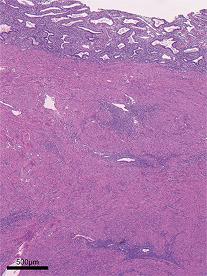
Adenomyosis
Secretory phase Age 42 - Whole
3D imaging and continuous tomographic image of adenomyosis in secretory phase.
Red object shows 3D structures of the direct invasion of the endometrial gland into the myometrium. Yellow objects show the ectopic endometrial glands which lengthened thin branches and formed a lesion like an ant colony within the myometrium. Gray object shows the eutopic endometrial glands.
3D reconstitution of adenomyosis imaged by light-sheet microscopy.
Red/yellow/gray: cytokeratin 7, Blue: autofluorescence